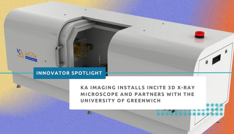
(Waterloo, 24 April 2023) – X-ray equipment manufacturer KA Imaging has announced a new install of the inCiTe™ 3D X-ray Microscope as part of a strategic partnership with the University of Greenwich, United Kingdom. The technology, first of its kind available in UK/Europe, will be used in research and knowledge transfer projects led by Professor Gianluca Tozzi.
The inCiTe™ 3D X-ray Microscope is the first commercial system that utilizes BrillianSe™, a patented high spatial resolution amorphous selenium (a-Se) detector exclusively developed by KA Imaging Inc.
The high spatial resolution and detection efficiency of the BrillianSe™ X-ray detector enable rapid phase-contrast imaging and conventional micro-CT in a portable benchtop system. Thanks to such phase-contrast technology, the inCiTe™ 3D X-ray Microscope provides superior contrast, and better visualization of low-density materials.
The new inCiTe™ unit will support joint research efforts aimed at further advancing fast phase-contrast tomography of soft tissues and biomaterials in the laboratory.
“inCiTe’s phase contrast technology is particularly beneficial for objects with low conventional absorption contrast, such as the soft biological tissues studied by Professor Tozzi,” explained Chris Scott, Technical Lead at KA Imaging. “We had very promising initial results with some samples we processed in inCiTe, where bone and soft tissue can be better visualized thanks to phase-contrast,” he continued.
Professor Gianluca Tozzi is Professor of Industrial Engineering at the Faculty of Engineering and Science, University of Greenwich, United Kingdom. His research is devoted to the understanding of structure/function of biological tissues and biomaterials using experimental techniques such as such as digital volume correlation (DVC) based on X-ray computed tomography, aimed at informing product development for industry as well as clinical practice for traumatic and pathological conditions. “I am very excited by the unique capability of the inCiTe 3D X-ray Microscope and potential for further technological development of phase-contrast tomography, within this partnership with KA Imaging,” said Professor Tozzi. “,” said the Professor.


